CDV Fusion glycoprotein F0 Mouse mAb (一抗) - WB,ELISA | Bioss
货号:bsm-49050M
产品详情
相关标记
相关产品
相关文献
常见问题
概述
产品编号
bsm-49050M
英文名称
CDV Fusion glycoprotein F0 Mouse mAb
中文名称
犬瘟热病毒融合糖蛋白F0单克隆抗体
英文别名
Canine Distemper Virus Fusion Glycoprotein F0; Protein F;
抗体来源
Mouse
免疫原
Recombinant Canine distemper virus Fusion glycoprotein F0: 136-600/662
亚型
IgG
性状
Size : 50ul/100ul/200ul
Liquid
Size : 200ug (PBS only)
Lyophilized
Note: Centrifuge tubes before opening. Reconstitute the lyophilized product in distilled water. Optimal concentration should be determined by the end user.
Liquid
Size : 200ug (PBS only)
Lyophilized
Note: Centrifuge tubes before opening. Reconstitute the lyophilized product in distilled water. Optimal concentration should be determined by the end user.
纯化方法
affinity purified by Protein A
克隆类型
Monoclonal
克隆号
4F2
理论分子量
73 kDa
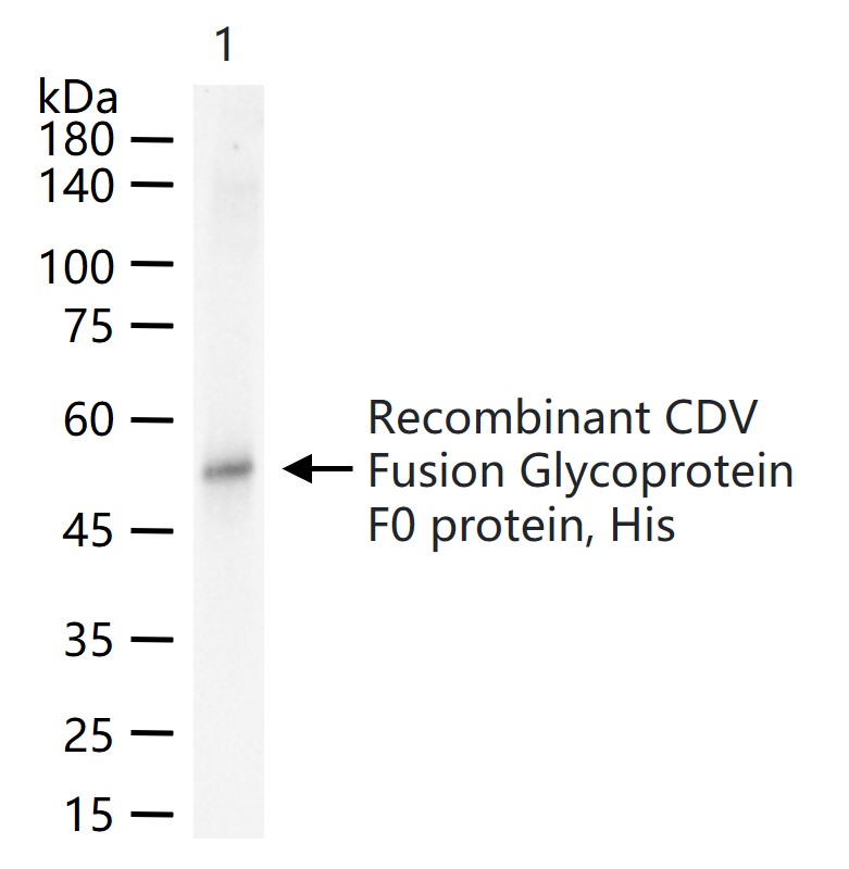
检测分子量
52 kDa
浓度
1mg/ml
储存液
Size : 50ul/100ul/200ul
0.01M TBS (pH7.4) with 1% BSA, 0.02% Proclin300 and 50% Glycerol.
Size : 200ug (PBS only)
0.01M PBS
0.01M TBS (pH7.4) with 1% BSA, 0.02% Proclin300 and 50% Glycerol.
Size : 200ug (PBS only)
0.01M PBS
保存条件
Shipped at 4℃. Store at -20℃ for one year. Avoid repeated freeze/thaw cycles.
注意事项
This product as supplied is intended for research use only, not for use in human, therapeutic or diagnostic applications.
背景资料
Class I viral fusion protein. Under the current model, the protein has at least 3 conformational states: pre-fusion native state, pre-hairpin intermediate state, and post-fusion hairpin state. During viral and plasma cell membrane fusion, the heptad repeat (HR) regions assume a trimer-of-hairpins structure, positioning the fusion peptide in close proximity to the C-terminal region of the ectodomain. The formation of this structure appears to drive apposition and subsequent fusion of viral and plasma cell membranes. Directs fusion of viral and cellular membranes leading to delivery of the nucleocapsid into the cytoplasm. This fusion is pH independent and occurs directly at the outer cell membrane. The trimer of F1-F2 (F protein) probably interacts with H at the virion surface. Upon HN binding to its cellular receptor, the hydrophobic fusion peptide is unmasked and interacts with the cellular membrane, inducing the fusion between cell and virion membranes. Later in infection, F proteins expressed at the plasma membrane of infected cells could mediate fusion with adjacent cells to form syncytia, a cytopathic effect that could lead to tissue necrosis
产品应用
| 应用 | 已检合格种属 | 预测种属 | 推荐稀释比例 |
|---|---|---|---|
| WB | CDV | 1:500-2000 | |
| ELISA | CDV | 1:5000-10000 |
交叉反应
交叉反应: CDV
相关产品
暂无相关产品
靶标
基因名
F
蛋白名
Fusion glycoprotein F0
同靶标产品
相关文献
提示: 发表研究结果有使用 bsm-49050M 时请让我们知道,以便我们可以引用参考文章。作为回馈,资料提供者将获得我们送上的小礼品。